


Press Release


From Star Charts to Microscopes: New Tool Solves Imaging Challenges with The Help of Astronomy
Finding the same microscopic field of view twice can be surprisingly difficult. A sample may be viewed at a different magnification, moved to another microscope or imaged again after further processing. Each change can alter how the sample looks, making it difficult for researchers to manually find the original area again. Researchers have now found a solution to this problem by borrowing an idea from astronomy.
Researchers from Turku Bioscience Centre and Åbo Akademi University, with collaborators from NOVA University Lisbon in Portugal and Lund University in Sweden have developed NucleiSky, an open-source tool that helps microscopes recognise and return to the same area of a biological sample. The tool treats clusters of cell nuclei as constellations that indicate where an image belongs.
Astronomers can identify an unknown region of the sky by comparing the arrangement of its stars with a star map. Similarly, NucleiSky converts the positions of cell nuclei into a constellation and searches for the same pattern in a larger microscopy image.
“Some of the best ideas can come from outside our own field. NucleiSky shows how methods developed for astronomy can solve practical problems in microscopy,” said Professor Guillaume Jacquemet.
Because NucleiSky uses the arrangement of nuclei rather than the overall appearance of an image, it can match fields of view captured at different magnifications, with different imaging methods or on different microscopes. Under favourable conditions, researchers have shown that as few as five nuclei were enough to identify the correct location.
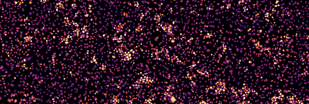
To test NucleiSky in a smart microscopy application, the researchers first imaged an area of a cell sample with one microscope and then transferred the sample to a second microscope using a different imaging modality. The second microscope, equipped with the smart microscopy workflow, automatically scanned the sample at low magnification, recognised the original constellation of nuclei, and navigated back to the same area. It then collected a higher-resolution image of that area using the new imaging modality.
“With NucleiSky, a microscope can recognise a field of view and return to it automatically. This opens up new possibilities for smart microscopy,” said doctoral researcher Iván Hidalgo-Cenalmor, the first author of the study.
NucleiSky could make complex imaging experiments faster and easier to reproduce by reducing the need for manual searching, special grids or artificial markers. It could also help enable future smart microscope workflows that explore samples, identify interesting areas and automatically return to them for detailed imaging.
The study on NucleiSky was published in the Journal of Cell Science and can be read here: https://doi.org/10.1242/jcs.265152
NucleiSky was developed within the framework of InFLAMES. The InFLAMES Research Flagship Centre is a joint initiative of the University of Turku and Åbo Akademi University. It brings together researchers working to develop new diagnostic and therapeutic tools for precision medicine.
For more information, please contact:
Guillaume Jacquemet, Professor of Bioimaging, Åbo Akademi University
Email: guillaume.jacquemet@abo.fi
Telephone: +358 50 323 5606

You might also be interested in
-
The Research Council of Finland funds cutting edge research at Åbo Akademi University with €4.5 million
Researchers at Åbo Akademi University have been granted nearly €4.5 million by the Research Council of Finland. The funding supports high-level research in fields such…

-

Euro-BioImaging in Turku secures major EU funding – Turku emerging as a European capital of biological and biomedical imaging
Euro‑BioImaging, the European research infrastructure network for biological and biomedical imaging headquartered in Turku, has been awarded substantial funding through the EU’s research and innovation…
-
Guillaume Jacquemet appointed to the prestigious K. Albin Johansson Research Professorship
Guillaume Jacquemet, Associate Professor of Cell Biology at Åbo Akademi University and Academy of Finland Research Fellow, and member of the InFLAMES Flagship, has been…